Shoulder Tendon Anatomy Diagram - 1 : Name the arteries and the nerves that supply shoulder joint.. Name the arteries and the nerves that supply shoulder joint. Shoulder joint anatomy skeletal system cartilages ligaments. Upper limb trauma programme injuries. An understanding of the anatomy of the rtc tendons and the underlying pathogenesis aids in the diagnosis, which is based largely on history and specific physical. Sechrest, md narrates an animated tutorial on the basic anatomy of the shoulder.
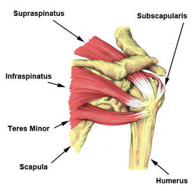
The most common shoulder injuries involve the muscles, ligaments, cartilage, and tendons. There are 10 muscles and 11 shoulder tendons related to shoulder mobility. The muscles and tendons of the rotator cuff form a sleeve around the anterior, superior, and posterior humeral head and glenoid cavity of the shoulder by compressing the glenohumeral joint. Draw labelled diagram showing the relations of shoulder joint. Specifically, the four rotator cuff muscles include the following
Causes And Treatment For Rotator Cuff Tears from www.advancephysiotherapy.co.nz The muscles and tendons of the rotator cuff form a sleeve around the anterior, superior, and posterior humeral head and glenoid cavity of the shoulder by compressing the glenohumeral joint. Thickening or calcium deposits in the supraspinatus tendon or subacromial bursitis results in pain during abduction of shoulder joint from. The shoulder has about eight muscles that attach to the scapula, humerus, and clavicle. Anterior graphic of the shoulder. Draw labelled diagram showing the relations of shoulder joint. It is constructed in such a way that we can move the arms to. For more anatomy content please follow us and visit our website: Shoulder joint anatomy skeletal system cartilages ligaments.
The subacromial bursa lies on the top portion of the supraspinatus tendon.
Extraocular muscles of left eye. Specifically, the four rotator cuff muscles include the following This mri shoulder axial cross sectional anatomy tool is absolutely free to use. An image depicting shoulder anatomy can be seen below. The shoulder joint offers a fuller range of motion than any other joint in the body. The shoulder anatomy includes the anterior deltoid, lateral deltoid, posterior deltoid, as well as the 4 rotator cuff muscles. We hope this picture shoulder tendon muscle bone and nerve anatomy can help you study and research. Name the arteries and the nerves that supply shoulder joint. The shoulder has about eight muscles that attach to the scapula, humerus, and clavicle. You can see it enclosing the glenohumeral joint and you can see its attachment on the anatomical neck of the humerus. Upper limb trauma programme injuries. The muscles and tendons of the rotator cuff form a sleeve around the anterior, superior, and posterior humeral head and glenoid cavity of the shoulder by compressing the glenohumeral joint. Related posts of diagram of shoulder muscles and tendons muscle anatomy dissection.
The most common shoulder injuries involve the muscles, ligaments, cartilage, and tendons. Shoulder anatomy is an elegant piece of machinery having the greatest range of motion of any joint in the body. The shoulder has about eight muscles that attach to the scapula, humerus, and clavicle. Shoulder radiology & anatomy at usuhs.mil. Normal anatomy, variants and checklist.
1 from An image depicting shoulder anatomy can be seen below. The shoulder joint (glenohumeral joint) is a ball and socket joint between the scapula and the humerus. The shoulder has about eight muscles that attach to the scapula, humerus, and clavicle. Sechrest, md narrates an animated tutorial on the basic anatomy of the shoulder. You can see these areas marked with an x in the shoulder anatomy diagram above. Ligaments are soft tissue structures that connect bones to bones. Shoulder joint anatomy skeletal system cartilages ligaments. There are several important ligaments in the shoulder.
Muscles allow us to move by pulling on bones.
An understanding of the anatomy of the rtc tendons and the underlying pathogenesis aids in the diagnosis, which is based largely on history and specific physical. Muscles allow us to move by pulling on bones. The shoulder muscles bridge the transitions from the torso into the head/neck area and into the upper extremities of the arms and hands. Shoulder muscles and shoulder tendons. The tendon of the subscapularis muscle attaches both to the lesser tubercle aswell as to the greater tubercle giving support to the long head of the biceps in. It is constructed in such a way that we can move the arms to. Draw labelled diagram showing the relations of shoulder joint. .anatomy, shoulder joints and muscles, shoulder structure anatomy, shoulder tendon anatomy, shoulder tendons ligaments, human muscles, bones in shoulder, ligaments of the related posts of diagram of shoulder muscles and tendons. Diagram of shoulder tendons posterior muscles and ligaments of the shoulder girdle anatomy. The rotator cuff andwhere the bicep tendon meets the shoulder. There are several important ligaments in the shoulder. Lining the fibrous membrane, you've got the synovial membrane. For more anatomy content please follow us and visit our website:
These muscles form the outer shape of the shoulder and underarm. Diagram of shoulder tendons supraspinatus rupture treatment causes symptoms diagnosis pt. Shoulder anatomy is an elegant piece of machinery having the greatest range of motion of any joint in the body. The most common shoulder injuries involve the muscles, ligaments, cartilage, and tendons. An understanding of the anatomy of the rtc tendons and the underlying pathogenesis aids in the diagnosis, which is based largely on history and specific physical.
Rotator Cuff Tear Wikem from wikem.org The rotator cuff andwhere the bicep tendon meets the shoulder. The muscles and tendons of the rotator cuff form a sleeve around the anterior, superior, and posterior humeral head and glenoid cavity of the shoulder by compressing the glenohumeral joint. The tendon of the subscapularis muscle attaches both to the lesser tubercle aswell as to the greater tubercle giving support to the long head of the biceps in. Related online courses on physioplus. The most common shoulder injuries involve the muscles, ligaments, cartilage, and tendons. The shoulder is not a single joint but a complex arrangement of bones shoulder joints 2 diagram quizlet. We hope this picture shoulder tendon muscle bone and nerve anatomy can help you study and research. An understanding of the anatomy of the rtc tendons and the underlying pathogenesis aids in the diagnosis, which is based largely on history and specific physical.
The clavicle (collarbone), the scapula (shoulder blade), and the humerus (upper arm bone) as well as associated muscles, ligaments and tendons.
Anterior graphic of the shoulder. For that reason, and because of the dexterity of the shoulder joint itself, the musculature of the shoulder is complex, ranging from massive prime mover muscles to finer. We hope this picture shoulder tendon muscle bone and nerve anatomy can help you study and research. This page is about shoulder tendon anatomy diagram,contains muscles attachment of rotator cuff muscle,upper extremity occupational therapy 205 with teresa at tufts university,soft tissues of the shoulder,biceps and triceps tendon rupture and more. It reduces wear and tear in this article, we shall look at the anatomy of the shoulder joint and its important clinical. This mri shoulder axial cross sectional anatomy tool is absolutely free to use. Shoulder muscles and shoulder tendons. There are 10 muscles and 11 shoulder tendons related to shoulder mobility. Name the arteries and the nerves that supply shoulder joint. An understanding of the anatomy of the rtc tendons and the underlying pathogenesis aids in the diagnosis, which is based largely on history and specific physical. Shoulder radiology & anatomy at usuhs.mil. Normal anatomy, variants and checklist. For more anatomy content please follow us and visit our website:
Thickening or calcium deposits in the supraspinatus tendon or subacromial bursitis results in pain during abduction of shoulder joint from shoulder anatomy diagram. The most important extrinsic soft tissues are the supraspinatus tendon superiorly, infraspinatus posteriorly and subscapularis anteriorly (fig.


0 Comments